


Ultrasound Medical Imaging has revolutionized the field of healthcare, enabling physicians to diagnose and monitor diseases safely and noninvasively. Let’s dive into the fascinating world of ultrasound technology and its impact on medical imaging.
The history of medical ultrasound dates back to the early 20th century, when scientists experimented with sound waves and their ability to penetrate tissue. In the 1960s, significant advances occurred as imaging techniques became more refined and practical for medical use.
In a Nutshell
- Ultrasound is a widely used medical imaging technique that uses high frequency sound waves to produce real time images of internal body structures.
- It is noninvasive, safe and does not involve exposure to ionizing radiation, making it the preferred choice for a variety of diagnostic purposes.
- Ultrasound Medical Imaging is commonly used to visualize organs such as the heart, liver, kidneys and reproductive organs to detect abnormalities or monitor disease.
- It is also used in obstetrics to monitor fetal development and during pregnancy to detect any complications or abnormalities.
- Ultrasound Medical Imaging can help guide medical procedures, such as biopsies and injections, by providing real time images of the target area.
- It is also an accessible and cost effective imaging modality, making it especially valuable in resource poor settings or areas with limited medical infrastructure.
Today, ultrasound technology has progressed by leaps and bounds thanks to modern advances. High resolution imaging, improved transducer design and sophisticated software have made ultrasound an indispensable tool for medical professionals. Its versatility makes it possible to obtain detailed images of various parts of the body, such as the heart, abdomen and reproductive system.
But how does ultrasound technology work? In essence, ultrasound uses sound waves to create images of internal structures. These waves, emitted by a probe called a transducer, travel through the body and bounce back when they encounter various tissues. The ultrasound machine translates the returned waves into images that provide valuable information about the patient’s condition.
Ultrasonography is arguably the most important advance in obstetrics and gynecology of the 20th century.
Stuart Campbell
Not all ultrasound scans are the same. Different types of ultrasound are used depending on the specific medical situation. For example, obstetric ultrasounds are often used during prenatal care, while echocardiograms provide detailed images of the structure and function of the heart. Doppler ultrasound, on the other hand, evaluates blood flow through blood vessels, which helps diagnose conditions such as deep vein thrombosis.
Ultrasound Medical Imaging continues to contribute significantly to the field of medicine, enabling accurate diagnoses, guiding surgical interventions and monitoring the progress of treatments. Thanks to its noninvasive nature and outstanding imaging capabilities, ultrasound has become an integral part of healthcare, ensuring safer and more effective patient care.
How Ultrasound Medical Imaging Is Used
Ultrasound plays a crucial role in medical imaging, providing valuable information about the internal structures of the human body. In this article we will explain the principle of operation of medical ultrasound, the generation and reception of ultrasonic waves and the formation of an ultrasound image. We will also discuss the different types of ultrasound imaging techniques used in medical practice.
Ultrasound Medical Imaging is based on sound waves with frequencies above the audible range, usually between 1 and 20 megahertz. To generate these waves, a transducer, a device that emits and receives ultrasound waves, is placed on the patient’s skin. The transducer contains piezoelectric crystals that convert electrical energy into sound waves and vice versa.
As the ultrasound waves penetrate the body, they encounter various tissues, causing some of the waves to be reflected back to the transducer. By analyzing the reflected waves, a computer can create a two dimensional image of the internal structures.
There are several types of ultrasound imaging techniques. Grayscale ultrasound is the most common and provides high resolution images that show the boundaries and textures of organs and tissues. Doppler ultrasound, on the other hand, makes it possible to visualize blood flow in real time by analyzing the frequency changes of the reflected waves.
In recent years, 3D and 4D ultrasound techniques have gained popularity. These techniques create three dimensional images of fetal development and provide detailed views of growing organs and facial features.
In conclusion, ultrasound is a powerful medical imaging tool that enables healthcare professionals to visualize and diagnose a wide range of conditions. Understanding the principles of operation and the various imaging techniques can help us appreciate the important impact of ultrasound on modern medicine.
Key Components of an Ultrasound Medical Imaging System
These components work together seamlessly to provide accurate and detailed results. Let’s take a closer look at the essential elements that make up an ultrasound system.
First, we have the ultrasound probe, also known as the transducer. This device is responsible for emitting and receiving the sound waves. It is made up of multiple crystals that generate sound waves when electrical impulses are applied to them. The probe is designed to be placed on the patient’s body and moved to capture images of the desired area. Different probe designs and frequencies are available to meet different imaging needs.
Advertisement

Next, we have the central processing unit (CPU), the brain of the ultrasound system. The CPU processes the signals received from the probe and converts them into high resolution images. It controls the various functions of the system, such as adjusting settings, optimizing image quality and managing data storage.
Finally, we come to the display, where the images produced by the ultrasound system are visualized. The display plays a crucial role in providing a clear and detailed view of the scanned area. Modern ultrasound scanners have high resolution monitors that enable healthcare professionals to examine the images accurately.
Advanced technologies such as color Doppler and 3D/4D imaging can also be displayed on the monitor, providing additional diagnostic information.

In summary, an ultrasound system consists of a probe, a central processing unit (CPU) and a display. These components work together to produce high quality images that aid in the diagnosis and treatment of various medical conditions. With continuous technological advances, medical ultrasound has become an indispensable tool in modern healthcare.
Benefits of Ultrasound Medical Imaging in Diagnosis
Ultrasound Medical Imaging has revolutionized the field of diagnostics and offers a number of advantages that make it the preferred choice of healthcare professionals. One of the most significant advantages is their non invasive nature, which provides patients with a sense of security during the imaging process. Unlike other techniques such as MRI or CT, ultrasound does not require incisions or radiation exposure, ensuring minimal discomfort and reduced risk.
In addition, ultrasound is considered safe for patients of all ages, including pregnant women and children, due to its non ionizing nature. This makes it an ideal choice for routine check ups and monitoring of various conditions over time, without worrying about exposure to harmful radiation.
Another key advantage of ultrasound is its versatility and wide application in various fields of medicine. It can be used to visualize and examine different parts of the body, such as organs, muscles, tendons and blood vessels. In addition, ultrasound helps to detect and diagnose numerous medical conditions, from heart disease and gallstones to liver disorders and cancerous tumors.
In obstetrics and gynecology, ultrasound plays a key role in monitoring fetal development, assessing maternal health and diagnosing potential complications. It is also widely used in cardiology, urology and orthopedics, among other specialties, to help make accurate and timely diagnoses.
In conclusion, medical ultrasound offers important advantages in the diagnostic field. Its noninvasive nature provides patients with a sense of security, while the absence of radiation makes it safe for a wide range of individuals. In addition, its versatility and ability to examine various body parts and conditions make it an invaluable tool in the medical field. With these numerous advantages, ultrasound continues to contribute significantly to the accurate and effective diagnosis of medical conditions.
Practical Use Cases of Ultrasound Medical Imaging
In the realm of medical diagnostics, Ultrasound Medical Imaging stands as a cornerstone technology, offering versatile and essential insights across various specialties. This non invasive imaging technique has transformed healthcare, providing critical information in real time and aiding in early detection, diagnosis, and treatment planning. From obstetrics and cardiology to musculoskeletal and abdominal imaging, ultrasound’s applications are as diverse as they are impactful. Let’s explore some of the practical use cases of Ultrasound Medical Imaging, highlighting its significance in different medical fields.
1. Obstetrics and Gynecology
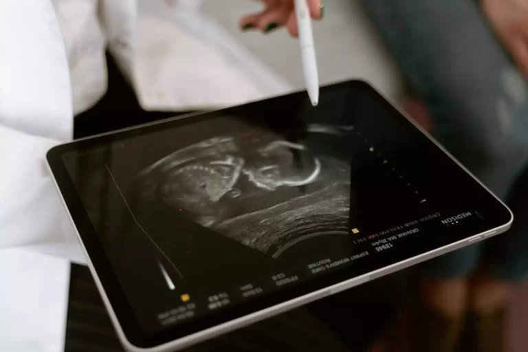
Prenatal ultrasounds are routine procedures performed during pregnancy to assess the development and well being of the fetus. Using ultrasound, healthcare professionals can monitor fetal growth, determine the position of the baby and detect possible abnormalities. By visualizing the fetus in real time, Ultrasound Medical Imaging allows early detection of conditions such as neural tube defects, cardiac abnormalities and certain genetic disorders. In addition, prenatal ultrasounds allow expectant parents to bond with the fetus by providing images and videos of the baby.

Ultrasound Medical Imaging also plays a crucial role in diagnosing gynecological problems in women. It is commonly used to investigate conditions such as pelvic pain, abnormal uterine bleeding and ovarian cysts. Using transvaginal or transabdominal ultrasound techniques, healthcare professionals can visualize the reproductive organs and identify any abnormalities or structural changes. In case of suspected tumors or cancers, ultrasound guided biopsies can be performed to obtain tissue samples for further analysis. Early detection of gynecological problems by ultrasound allows for rapid intervention and improved patient outcomes.
2. Cardiology
Ultrasound Medical Imaging has also become an invaluable tool in cardiology, as it facilitates noninvasive evaluation of the structure and function of the heart. The following are two outstanding cases of the use of ultrasound in this field:
- Echocardiograms: Echocardiograms, also known as cardiac ultrasounds, are used to evaluate the structure and function of the heart. This noninvasive imaging technique allows cardiologists to visualize the heart chambers, valves and major blood vessels. By analyzing the echoes produced by ultrasound waves bouncing off cardiac tissues, healthcare professionals can assess the heart’s pumping ability and detect any abnormalities, such as valvular disorders, congenital heart defects or signs of heart failure. Echocardiograms are vital for diagnosing and monitoring various cardiovascular conditions, guiding therapeutic decisions and evaluating the effectiveness of interventions.
- Stress echocardiograms: Stress echocardiograms combine ultrasound with physical exertion to assess the heart’s response to exercise or medications that mimic the effects of exercise. This test helps identify abnormalities in blood flow to the heart muscle, detect areas of reduced function and determine if there are any signs of coronary artery disease. By monitoring the heart’s response under stress, stress echocardiography helps diagnose conditions such as blocked or narrowed arteries, heart valve abnormalities and heart muscle injury. This information helps cardiologists formulate appropriate treatment plans and stratify risk.
3. Musculoskeletal
Ultrasound Medical Imaging plays a crucial role in the detection and diagnosis of tendon, muscle and joint problems. Using high frequency sound waves, ultrasound can create detailed images of the body’s soft tissues, allowing healthcare professionals to identify abnormalities and provide accurate treatment plans.
One of the main advantages of ultrasound in musculoskeletal imaging is its ability to guide injections for arthritis or inflammation. This technique ensures precise placement of medication in the affected area, maximizing the effectiveness of treatment. By visualizing the exact location of the problem, ultrasound helps healthcare professionals deliver targeted and effective care.
In addition to its practical applications, Ultrasound Medical Imaging offers several advantages to patients. Compared to other imaging methods, such as MRI or CT, ultrasound is noninvasive and does not involve radiation exposure. It is a safe and painless procedure that can be performed in real time, providing immediate information to both the patient and the healthcare professional.
Advertisement
Ultrasound Medical Imaging has revolutionized the way musculoskeletal conditions are diagnosed and treated. Its versatility, accuracy and safety make it an indispensable tool for healthcare professionals. Whether you are suffering from joint pain, muscle strains or tendon injuries, ultrasound can help your healthcare professional identify the cause of your problems and develop an effective treatment plan tailored to your specific needs.
Don’t let musculoskeletal problems hinder your quality of life. Explore the advantages of ultrasound medical imaging and take control of your health today.
4. Abdominal Imaging
Ultrasound, an essential tool in medical imaging, plays a crucial role in the examination of various abdominal organs. It provides a safe and noninvasive method for visualizing internal structures such as the liver, kidneys and gallbladder, among others. This advanced imaging technique has revolutionized the diagnosis and treatment of abdominal conditions.

Abdominal Ultrasound Medical Imaging is especially useful in diagnosing the underlying causes of abdominal pain. By using sound waves to create real time images, physicians can assess the condition of organs and detect any abnormalities or potential problems. For example, ultrasound can detect liver disease such as cirrhosis or fatty liver, allowing for early intervention and treatment.
In addition to the liver, ultrasound is invaluable in evaluating the kidneys. Whether assessing kidney function, detecting kidney stones or identifying possible tumors, ultrasound helps to get a complete picture of kidney health.
In addition, Ultrasound Medical Imaging can evaluate the gallbladder, an organ often associated with conditions such as gallstones or inflammation. By visualizing the gallbladder, ultrasound allows for quick and accurate diagnosis, enabling physicians to recommend appropriate treatment plans promptly.
Overall, ultrasound as a medical imaging technique offers significant advantages for examining abdominal organs, diagnosing abdominal pain and facilitating healthcare decision making. Its noninvasive nature, coupled with its ability to provide real time imaging, makes it a vital tool in the medical field. Whether it is monitoring organ function, identifying abnormalities or assisting in the treatment of abdominal conditions, Ultrasound Medical Imaging continues to contribute greatly to patient care and treatment outcomes.
Risks and Limitations of Ultrasound Medical Imaging
Ultrasound Medical Imaging has revolutionized medical diagnostics, providing physicians with a noninvasive and painless way to visualize internal body structures. Although generally safe and widely used, there are some risks and limitations associated with this medical imaging technique.
One of the potential risks of ultrasound is radiation exposure. Unlike other imaging methods, such as X-rays or computed tomography, ultrasound does not use ionizing radiation, making it safer in this regard. However, overexposure to ultrasound waves for prolonged periods of time can cause an increase in tissue temperature, with consequent adverse effects.
Another limitation of Ultrasound Medical Imaging is the possibility of misdiagnosis due to image quality. The quality of the ultrasound image is highly dependent on factors such as the patient’s body composition, the presence of gas or bone that may obstruct the sound waves, and the skill level of the operator. In some cases, these factors can compromise image quality and lead to incorrect or inconclusive diagnoses.
In addition, Ultrasound Medical Imaging may have limitations in imaging certain parts of the body. For example, ultrasound waves do not penetrate bone or air, so imaging the brain or lungs may be difficult. In addition, the size of the patient may also be a limitation, as excess body fat or a large build may hinder the effectiveness of ultrasound.
In addition, the skill level of the operator plays a crucial role in the accuracy of the ultrasound. Interpretation of ultrasound images and the ability to detect subtle abnormalities require specialized training and expertise. Inexperienced or inadequately trained operators may have difficulty obtaining accurate results, which can lead to misdiagnosis or missed diagnoses.
In conclusion, although Ultrasound Medical Imaging is a valuable tool for medical diagnosis, it is important to be aware of its risks and limitations. Understanding these factors can help healthcare professionals make informed decisions and ensure the optimal use of ultrasound in patient care.
Wrap Up
Medical imaging is advancing rapidly, driven by technological advances and new trends. Of particular note is the integration of artificial intelligence (AI) into Ultrasound Medical Imaging. AI algorithms can analyze ultrasound images with remarkable accuracy, helping healthcare professionals diagnose diseases and detect abnormalities more accurately.
Another important trend is the rise of portable ultrasound scanners. These compact, easy to use devices allow medical professionals to perform point of care ultrasound scans, providing real time images without the need for large fixed equipment. This portability opens up new possibilities for remote and rural healthcare, as well as for emergency situations where immediate imaging is crucial.
In research, therapeutic Ultrasound Medical Imaging is gaining prominence. This innovative technique uses focused ultrasound waves to treat a variety of medical conditions, such as tumors, heart disease and neurological disorders. By directing ultrasound energy to specific targets in the body, therapeutic ultrasound can revolutionize treatment options and improve patient outcomes.
Ultrasound Medical Imaging is also being explored in combination with other imaging modalities. By integrating ultrasound with techniques such as computed tomography (CT) or magnetic resonance imaging (MRI), healthcare professionals can obtain more complete and detailed images, enabling more accurate diagnoses and treatment planning.
As technology advances and research progresses, the future of medical ultrasound looks bright. With advances in artificial intelligence improving diagnostic capabilities, portable devices increasing accessibility, and new areas of research such as therapeutic ultrasound and combinations with other imaging modalities, the field is poised to have an even greater impact on healthcare. Watch for developments in ultrasound and medical imaging that will revolutionize the way diseases are diagnosed and treated.
FAQs

Ultrasound is a medical imaging technique that uses high frequency sound waves to create images of the inside of the body.
Ultrasound Medical Imaging works by transmitting high frequency sound waves into the body. These waves bounce off internal tissues and organs, and the echoes are picked up by a transducer, which creates images based on the returned sound waves.
Ultrasound Medical Imaging is commonly used to visualize and diagnose conditions in various parts of the body, such as the abdomen, pelvis, heart, blood vessels and musculoskeletal system. It is also used during pregnancy to monitor the health and development of the fetus.
Yes, Ultrasound Medical Imaging is considered a safe imaging technique because it uses sound waves instead of ionizing radiation like X-rays. It is noninvasive and does not involve any potentially harmful radiation exposure.
In general, there are no known risks or side effects associated with Ultrasound Medical Imaging. It is a painless procedure that requires no special preparation, although occasionally mild discomfort may be experienced when the transducer is pressed against the body. However, as with any medical procedure, it is always recommended to consult a healthcare professional if you have specific questions.
Article sources
At Capital Maniacs, we are committed to providing accurate and reliable information on a wide range of financial topics. In order to achieve this, we rely on the use of primary sources and corroborated secondary sources to support the content of our articles.
Primary sources, such as financial statements and government reports, provide firsthand evidence of financial events and trends. By using primary sources, we are able to directly reference information provided by the organizations and individuals involved in these events.
Secondary sources, such as financial analysis and commentary, interpret and analyze primary sources. While these sources can be useful for providing context and background information, it is important to use corroborated sources in order to ensure the accuracy and reliability of the information we present.
We take pride in properly citing all of our sources, both primary and secondary, in order to give credit to the original authors and to allow our readers to verify the information for themselves. We appreciate your trust in our website and are committed to upholding the highest standards of financial journalism.
- Fda.gov – Ultrasound Imaging
- Mayoclinic.org – Ultrasound – Mayo Clinic
- Nibib.nih.gov – What is medical ultrasound?
- En.wikipedia.org – Medical ultrasound – Wikipedia
- Ncbi.nlm.nih.gov – Ultrasound – Medical Imaging Systems – NCBI Bookshelf
- Unlv.edu – Bachelor of Science in Comprehensive Medical Imaging
- Ncbi.nlm.nih.gov – Application of Ultrasound in Medicine – PMC